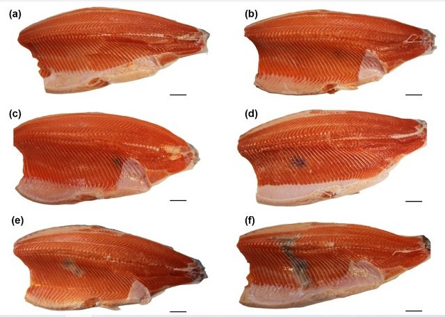
Imagine going to buy salmon fillets only to encounter unaesthetic dark spots that spoil their presentation. These imperfections, known as “melanized focal changes” (MFC), pose a significant challenge to the salmon industry, significantly impacting the market value of the fish.
Over 20% of Atlantic salmon fillets may have unattractive black and red spots, often measuring >1 cm, resulting in substantial financial losses. These spots are much more prevalent in farmed salmon than in the wild, and their causes are not well understood.
But what causes these dark discolorations?
Recognizing the need to clarify the biochemistry of these spots, Professor Turid Mørkøre from the Norwegian University of Life Sciences turned to two leaders in melanin biochemistry, Professor Kazumasa Wakamatsu and Professor Shosuke Ito from Fujita Health University, Japan.
The research team delved into this mystery, employing sophisticated chemical techniques to reveal the true nature of these discolorations. The results shed light on the fascinating interaction between inflammation, immune response, and pigment production in salmon, offering valuable insights to enhance the quality of salmon fillets.
The Problem of Spots on Salmon Fillets
In recent decades, focal discolorations on Atlantic salmon muscle fillets have become an increasing issue for the commercial cultivation of seafood, affecting a substantial portion of the fillets. The reasons for these spots are mysterious, though several plausible theories, such as viral infection and rib fractures, have been proposed. However, multiple different causes are likely. The spots are roughly classified as “red spots” or “black spots” and are also known as “red focal changes” and “melanized focal changes.”
However, a proper biochemical analysis of black spots had never been conducted, and the suggestion that they contained melanin was mainly based on Fontana Masson staining, which is only relatively specific for melanin. Intermediate forms between red and black spots have been found, leading to a shared concept that black spots generally derive from red spots.
Melanin Under the Microscope
Previous research hinted at the involvement of melanin in MFC but lacked concrete evidence.
Melanin is a large and irregular heteropolymer consisting of monomeric units derived from the enzymatic oxidation of the amino acid tyrosine, and there are several types of melanin. Professors Wakamatsu and Ito are melanin specialists who established a panel of biochemical assays to characterize multiple types of melanin for different species, ranging from human patients with melanoma to fossilized animals like dinosaurs.
Stay Always Informed
Join our communities to instantly receive the most important news, reports, and analysis from the aquaculture industry.
The study addressed this gap using a powerful duo of chemical methods: alkaline oxidation with hydrogen peroxide and hydrolysis with hydriodic acid. These techniques allowed researchers to accurately identify the type of melanin present in both MFC and red pigment regions, known as “red focal changes (RFC).”
The Results?
Researchers discovered that black spots contain a type of melanin called eumelanin, while red spots do not contain detectable melanin. The biochemical discontinuity between red and black spots supports that their pigments derive from different cellular origins—red blood cells and melanomacrophages (fish macrophages with dark pigment), respectively.
The findings were enlightening. Indeed, MFC contained high levels of eumelanin, the same dark pigment found in our hair. This confirms the suspected role of melanomacrophages, immune cells that accumulate melanin while cleaning cellular waste. In contrast, RFC showed only traces of eumelanin.
While melanin takes center stage, researchers suspect that other factors contribute to the red stain mystery. Certain fragments of oxidized proteins and remnants of the inflammatory process may also be at play. Further research is needed to fully understand their role.
The False Lead
The red pigment presented a different enigma. Chemical analysis failed to identify significant levels of eumelanin or “pheomelanin.” Therefore, the red color likely originates from something completely different—most likely bleeding or hemorrhaging within the muscle tissue.
While melanin played a central role in MFC, the story of RFC was not as clear. Researchers observed a correlation between the intensity of red pigment and the presence of certain melanogenic metabolites. These are intermediate molecules produced during melanin synthesis but do not necessarily form the final pigment itself.
Imitation Experiment
To better understand this, researchers conducted an “imitation experiment.” They exposed a mixture of salmon muscle tissue and DOPA (a precursor to melanin) to the enzyme tyrosinase, which plays a crucial role in melanin production. The resulting mixture exhibited a reddish color similar to RFC, suggesting that DOPAquinone and/or DOPAchrome, other intermediaries in the melanin pathway, could bind to salmon muscle proteins, contributing to the red coloration.
In essence, the study reveals that the origins of MFC and RFC lie in distinct chemical pathways, reflecting their different cellular origins. MFC, stemming from melanomacrophages, accumulates eumelanin as part of their immune response. RFC, on the other hand, likely involves bleeding and the binding of melanogenic metabolites to muscle proteins, without significant melanin formation.
It’s important to note that Wakamatsu et al.’s findings revealed no biochemical continuity between the pigments of red and black spots, supporting previous hypotheses based on histology that red spots are caused by bleeding, and black spots by local accumulations of melanomacrophages in chronic local immune reactions.
Melanomacrophages
Melanomacrophages are immune cells found only in ectothermic vertebrates, including fish, amphibians, and reptiles. By inference, Wakamatsu et al.’s new study implies that the black pigment in melanomacrophages is eumelanin, which had not been properly determined before.
Hemorrhages
Hemorrhages can have various causes, not all of which lead to chronic inflammation with melanomacrophages, and melanomacrophages can also accumulate for reasons other than bleeding. Therefore, the distinct cellular origin of red and black spots, supported by their biochemical discontinuity shown by Wakamatsu et al., implies that researchers should expect a variety of possible causes for different stains and not try to find a one-size-fits-all explanation.
Implications for the Salmon Industry
This study paves the way for better strategies to combat black spots. By understanding the biochemistry and progression of these discolorations, researchers and salmon farmers can develop specific solutions. This could involve optimizing cultivation conditions, improving fish health, or even manipulating melanin production pathways to minimize both the aesthetic imperfections of salmon fillets and potential health issues associated with inflammation. In this regard, a study by researchers at Nofima highlighted that the omega-3 fatty acid DHA inhibits the synthesis of the pigment melanin.
Professor Erling Koppang, a Norwegian expert on salmon fillet spots who did not participate in this study, explains: “Wakamatsu’s study is an important component in characterizing pigmented lesions in Atlantic salmon and aligns very well with that of our group. Previous identification of tyrosinase expression [a gene necessary for melanin production] in the black changes.”
“We now know with certainty that the end product is what we expected, which is important for advancing attempts to ban these lesions.”
Conclusion
The researchers conclude that from the study’s results, it can be inferred that the pigment in melanomacrophages (the source of MFC) is eumelanin derived from DOPA, which was anticipated but fills a crucial gap in understanding the immune system of primitive vertebrates.
They also recommend that to reduce RFC and MFC, future research should strive to learn how to prohibit bleeding and (other) inflammations in farmed salmon muscle tissue, possibly addressing handling injuries, high population density, nets, and/or reducing the pathogen load from eutrophied cage water.
Black stains on salmon are more than simple cosmetic imperfections; they represent a complex interaction of inflammation, immune response, and pigment production. This study sheds new light on these processes and offers hope for combating undesirable dark spots on salmon fillets.
The study has been funded by The Norwegian Seafood Research Fund.
Reference (open access)
Wakamatsu, Kazumasa, Johannes M. Dijkstra, Turid Mørkøre, and Shosuke Ito. 2023. “Eumelanin Detection in Melanized Focal Changes but Not in Red Focal Changes on Atlantic Salmon (Salmo salar) Fillets” International Journal of Molecular Sciences 24, no. 23: 16797. https://doi.org/10.3390/ijms242316797
Editor at the digital magazine AquaHoy. He holds a degree in Aquaculture Biology from the National University of Santa (UNS) and a Master’s degree in Science and Innovation Management from the Polytechnic University of Valencia, with postgraduate diplomas in Business Innovation and Innovation Management. He possesses extensive experience in the aquaculture and fisheries sector, having led the Fisheries Innovation Unit of the National Program for Innovation in Fisheries and Aquaculture (PNIPA). He has served as a senior consultant in technology watch, an innovation project formulator and advisor, and a lecturer at UNS. He is a member of the Peruvian College of Biologists and was recognized by the World Aquaculture Society (WAS) in 2016 for his contribution to aquaculture.


