Discolored spots sometimes appear on farmed fish fillets, such as salmon and trout. These red and black discolorations are a concern for both fish farmers and consumers.
A study led by scientists from the Norwegian University of Life Sciences, Pure Salmon Kaldnes, and the Norwegian Veterinary Institute examines discolorations, called melanized focal changes (MFC), in rainbow trout (Oncorhynchus mykiss) fillets and compares them to those found in Atlantic salmon (Salmo salar). While these discolorations are well-documented in Atlantic salmon, less is known about their prevalence and characteristics in rainbow trout.
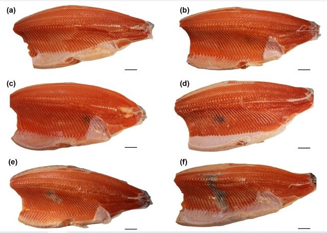
Discolored Fillets: A Concern for Fish Farmers
Up to 20% of Atlantic salmon fillets can be affected by MFC, causing significant economic losses. In contrast, MFC were much less frequent in rainbow trout, ranging from only 1.46% to 6.47% depending on the farm.
These discolorations are caused by melanomacrophages, pigment-producing cells that accumulate in the muscle. While these cells are harmless, they make the fillets less appealing to consumers.
The study focused on red focal changes (RFC) and melanized focal changes (MFC) found in the muscle tissue (fillets) of rainbow trout. These discolorations are similar to those observed in Atlantic salmon. While both species are commercially important, rainbow trout appears to have a much lower prevalence of these discolorations compared to salmon.
Researchers in Norway examined more than 1,200 rainbow trout from three different farms at the time of slaughter. They inspected the fillets for discolorations both visually (macroscopically) and under a microscope (histologically). Additionally, they looked for the presence of specific viruses that might be related to the discoloration.
Findings: Lower Prevalence and Different Pathology
The study found a much lower prevalence of MFC in rainbow trout compared to Atlantic salmon. Furthermore, the MFC in trout showed different characteristics. While both fish have melanomacrophages (cells that contribute to dark discoloration), trout showed more fibrosis (scarring) and muscle regeneration compared to salmon. Interestingly, viruses (piscine orthoreovirus 1, PRV-1) often associated with these discolorations in salmon were present in trout but not necessarily related to the discolored areas.
Researchers propose two possible reasons for the lower prevalence and different characteristics of MFC in rainbow trout:
- Defense Mechanisms: Rainbow trout may have stronger natural defenses compared to salmon, allowing them to better combat infections that contribute to MFC.
- Immune Response: The trout’s immune system might react differently to viral infections, leading to a less severe form of MFC compared to salmon.
Rainbow Trout May Be More Resilient
These findings suggest that rainbow trout develop a different type of MFC compared to Atlantic salmon. The lower prevalence and predominance of fibrosis and regeneration may indicate that rainbow trout are more resilient to the factors causing these discolorations. The presence of viruses without a clear connection to the lesions further supports this idea.
Stay Always Informed
Join our communities to instantly receive the most important news, reports, and analysis from the aquaculture industry.
Future Research: Understanding the Differences
The study also investigated rainbow trout for the presence of other viruses known to infect salmonid fish (salmon and trout). While some viruses were detected, they were not consistently associated with the observed discolorations.
The location of the discolorations in both salmon and trout was similar, suggesting a possible common trigger for the initial tissue damage.
More research is needed to explore the differences in healing responses between rainbow trout and Atlantic salmon.
More research is needed to understand the exact causes of MFC in rainbow trout and why they differ from those in salmon. This knowledge could help improve fish farming practices to minimize discoloration and ensure high-quality fish fillets for consumers.
Conclusion
“Our study provides insights into the prevalence and key histopathological characteristics of MFC in rainbow trout raised in a marine environment. The observation that most of these changes appear in the cranio-ventral area is consistent between rainbow trout and Atlantic salmon, suggesting a shared underlying cause,” the researchers conclude.
Additionally, the research provides valuable information about the differences between rainbow trout and Atlantic salmon regarding the discolorations of their fillets. Understanding these variations can contribute to improving fish health and enhancing seafood quality for consumers.
The study was partially funded by the Norwegian Seafood Research Fund.
Contact
Erling Olaf Koppang
Unit of Anatomy, Faculty of Veterinary Medicine, Norwegian University of Life Sciences
1433 Ås, Norway
Email: erling.o.koppang@nmbu.no
Reference (open access)
Bjørgen H, Brimsholm M, Lund M, Dahle MK, Rimstad E, Koppang EO (2024) Red and melanized focal changes in the white skeletal muscle of farmed rainbow trout Oncorhynchus mykiss. Dis Aquat Org 158:201-213. https://doi.org/10.3354/dao03797
Editor at the digital magazine AquaHoy. He holds a degree in Aquaculture Biology from the National University of Santa (UNS) and a Master’s degree in Science and Innovation Management from the Polytechnic University of Valencia, with postgraduate diplomas in Business Innovation and Innovation Management. He possesses extensive experience in the aquaculture and fisheries sector, having led the Fisheries Innovation Unit of the National Program for Innovation in Fisheries and Aquaculture (PNIPA). He has served as a senior consultant in technology watch, an innovation project formulator and advisor, and a lecturer at UNS. He is a member of the Peruvian College of Biologists and was recognized by the World Aquaculture Society (WAS) in 2016 for his contribution to aquaculture.

: Complete care guide for aquarium, eeding, breeding, and common illnesses")
: Technology, Design, and Profitability")
 2026: A Comprehensive Guide to Aquaculture 4.0 via AI and IoT Integration")