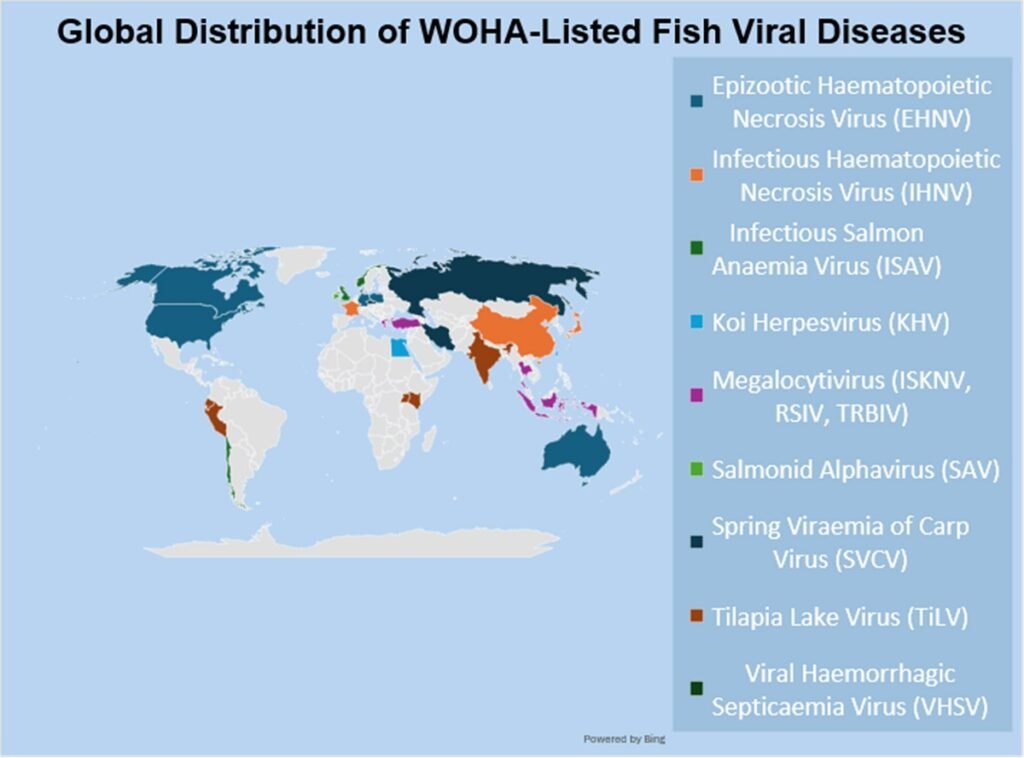
The accelerated growth of the aquaculture industry faces an invisible yet devastating enemy: viruses. Pathogens listed by the World Organisation for Animal Health (WOAH), such as Koi Herpesvirus (KHV) or Infectious Salmon Anemia Virus (ISAV), generate massive economic losses and restrict international trade.
This new meta-analysis, published in Reviews in Aquaculture, systematizes for the first time how these viruses “trace” their passage through the fish’s body, leaving microscopic scars on vital organs. Understanding these signals is not merely an academic exercise; it is the key to the industry’s survival.
- 1 Key Points
- 2 Study Methodology
- 3 The Spleen: Mirror of Immune Collapse
- 4 The Kidney: Osmotic Failure and Systemic Death
- 5 The Gills: The Broken Barrier
- 6 The Gastrointestinal Tract: The Intruder in Nutrition
- 7 The Challenge of Global Standardization
- 8 Conclusion: From Destruction to Resilience
- 9 Entradas relacionadas:
Key Points
- Sentinel Organs: The spleen and kidney act as the primary immunological “battlegrounds,” where necrosis and lymphoid depletion mark the collapse of the fish’s defensive system.
- Cellular Asphyxiation: Branchial viruses not only damage tissue; they trigger hyperplasia (excessive cell growth) that fuses lamellae, causing hypoxia and fatal osmoregulatory failure.
- The Impact of Age: Juvenile specimens exhibit disproportionately higher susceptibility, with more destructive lesions due to innate immune systems that are still developing.
- The Urge for Standardization: A critical gap exists in global aquaculture: the lack of harmonized lesion scoring systems, which prevents precise comparative diagnoses between laboratories.
Study Methodology
The team from the University of Arkansas at Pine Bluff conducted an exhaustive synthesis of decades of histopathological literature. The study compared lesion profiles across multiple fish families (salmonids, cyprinids, cichlids), cross-referencing data from both natural and experimental infections. Special emphasis was placed on Hematoxylin and Eosin (H&E) staining techniques and the identification of specific markers, such as syncytial cells and inclusion bodies.
The Spleen: Mirror of Immune Collapse
The spleen in teleost fish is the nerve center for immune response and hematopoiesis. When a virus like EHNV (Epizootic Hematopoietic Necrosis Virus) attacks, the spleen is the first to show signs of defeat: extensive splenic necrosis and drastic lymphoid depletion.
Interspecies Variations in the Spleen
| Virus | Affected Species | Characteristic Lesion |
| SVCV | Common Carp, Goldfish | Severe hemorrhage and dense inflammatory infiltrates. |
| ISAV | Atlantic Salmon | Congestion and hyperplastic melanomacrophage centers. |
| Megalocytivirus | Grouper, Sea Bream | Megalocytes with giant basophilic inclusions. |
Research highlights that, while findings are consistent, intensity varies according to water temperature. For instance, in rainbow trout, the severity of EHNV lesions is exacerbated by heat, suggesting that climate change could enhance viral lethality.
The Kidney: Osmotic Failure and Systemic Death
In fish, the kidney manages both filtration and blood cell production. Viruses such as IHNV (Infectious Hematopoietic Necrosis) transform renal tissue into a landscape of destruction: tubular necrosis and glomerular atrophy.
The consequences are twofold: the fish loses the ability to balance internal ions (osmoregulation), leading to interstitial edema, and the destruction of renal hematopoietic tissue induces profound anemia, leaving the animal defenseless against secondary infections. Viral inclusion bodies within the tubular epithelium act as diagnostic “fingerprints,” confirming active viral replication at the site.
The Gills: The Broken Barrier
The gills are perhaps the most vulnerable organ due to their direct contact with the aquatic environment. The study reveals a terrifying damage pattern termed lamellar fusion. Under attack from viruses like TiLV (Tilapia Lake Virus) or SAV (Salmon Alphavirus), branchial epithelial cells proliferate uncontrollably (hyperplasia) in a failed attempt at protection. This thickens the gill lamellae until they fuse, drastically reducing the oxygen exchange area. The fish literally suffocates in the water, even when oxygen levels are normal.
Stay Always Informed
Join our communities to instantly receive the most important news, reports, and analysis from the aquaculture industry.
The Gastrointestinal Tract: The Intruder in Nutrition
Often ignored in favor of more “visible” organs, the intestine suffers serious damage that undermines long-term vitality. Villous atrophy is the predominant lesion. When viruses like VHSV (Viral Hemorrhagic Septicemia Virus) infect the gut, villi shorten or fuse, eliminating the surface area required for nutrient absorption. Furthermore, the loss of the epithelial barrier allows opportunistic bacteria to invade the bloodstream, explaining why many viral outbreaks culminate in mixed bacterial septicemia.
The Challenge of Global Standardization
One of the most critical points discussed by Kurapati and his team is the diagnostic “Tower of Babel.” Currently, laboratories utilize different fixation methods, section thicknesses, and varied terminology to describe the same lesion. The lack of a universal lesion scoring system prevents scientists from rigorously comparing the susceptibility of different genetic fish varieties. Without this common metric, the development of vaccines and selective breeding programs for disease resistance becomes significantly slower and more costly.
Conclusion: From Destruction to Resilience
Analyzing the “histological landscapes” of these infections reveals a narrative of vulnerability but also of opportunity. These lesions are not just marks of destruction; they are anatomical maps indicating where we must bolster fish health. By integrating classical histopathology with modern tools like digital pathology and machine learning, aquaculture can shift from reactive to predictive. The future of sustainable fish farming depends on our ability to read these silent messages written in tissue before they escalate into economic disasters.
Contact
Yathish Ramena
University of Arkansas at Pine Bluff Center of Excellence
Pine Bluff, Arkansas, USA
Email: ramenay@uapb.edu
Reference (open access)
Kurapati, R. B., Ramena, G., Wanjala, H., Gudapati, S., & Ramena, Y. (2026). Viral Disease Histopathology in Aquaculture Finfish: Organ-Specific Pathological Changes and Diagnostic Insights, Referencing the World Organisation for Animal Health: A Review. Reviews in Aquaculture, 18, 270129. https://doi.org/10.1111/raq.70129
Editor at the digital magazine AquaHoy. He holds a degree in Aquaculture Biology from the National University of Santa (UNS) and a Master’s degree in Science and Innovation Management from the Polytechnic University of Valencia, with postgraduate diplomas in Business Innovation and Innovation Management. He possesses extensive experience in the aquaculture and fisheries sector, having led the Fisheries Innovation Unit of the National Program for Innovation in Fisheries and Aquaculture (PNIPA). He has served as a senior consultant in technology watch, an innovation project formulator and advisor, and a lecturer at UNS. He is a member of the Peruvian College of Biologists and was recognized by the World Aquaculture Society (WAS) in 2016 for his contribution to aquaculture.


: Technology, Design, and Profitability")