Despite scientific and technological advances, the main method of identifying and counting algae species is primarily through observation under a microscope, which is time-consuming and laborious.
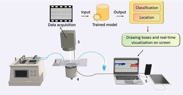
To monitor and determine the health status of marine microalgae in situ, hardware and software integration is needed to enable real-time algal imaging, data analysis, and implementation of machine learning algorithms to recognize and classify the cells of marine microalgae.
A team of researchers from Hainan University, Zhejiang University and The State University of New York at Stony Brook proposes a new database for the detection of marine microalgae and a set of detection methods.
The database includes images of six genera of microalgae (Pinnularia, Chlorella, Platymonas, Dunaliella salina, Isochrysis, and Symbiodinium) that are common in the ocean, and of the different developmental stages of the genera of microalgae.
Computer vision to identify microalgae
Object detection is a computer vision task that involves the location and identification of objects in an image or video.
The researchers applied computer vision algorithms for the detection of multiple objects and the assessment of the physiological state of marine microalgae.
“In this study, we trained one-stage object detection algorithms, including YOLOv5, YOLOv8, and TOOD, and two-stage object detection algorithms, including Faster-RCNN, Cascade-RCNN, and Dynamic-RCNN,” the researchers report.
Microalgae Image Database
The study authors established a database for the detection of marine microalgae in microscopy images, including collection data, microscopy images, etc.
The database contains 967 images of microalgae from six genera: Pinnularia, Chlorella, Platymonas, D. salina, Isochrysis, and Symbiodinium. In addition, they also collected images of Symbiodinium in different physiological states.
Mantente siempre informado
Stay Informed
Únete a nuestras comunidades para recibir al instante las noticias, informes y análisis más importantes del sector acuícola.
Join our communities to get instant access to the most important news, reports, and analysis from the aquaculture industry.
Conclusion
“The results showed that one-stage and two-stage object detection models can achieve high average accuracy, demonstrating the ability of computer vision in detecting multiple microalgae objects and providing basic data and models for detection in real-time from microalgae cells”, they conclude.
The study was funded by the Hainan Provincial Key Research and Development Program, the Hainan Provincial Higher Education Scientific Research Project Key Project, the Hainan Provincial Social Science Research Project, and the Hainan Provincial Natural Science Foundation.
Reference (open access):
Zhou, S., Jiang, J., Hong, X., Fu, P., & Yan, H. (2023). Vision meets algae: A novel way for microalgae recognization and health monitor. Frontiers in Marine Science.
Editor at the digital magazine AquaHoy. He holds a degree in Aquaculture Biology from the National University of Santa (UNS) and a Master’s degree in Science and Innovation Management from the Polytechnic University of Valencia, with postgraduate diplomas in Business Innovation and Innovation Management. He possesses extensive experience in the aquaculture and fisheries sector, having led the Fisheries Innovation Unit of the National Program for Innovation in Fisheries and Aquaculture (PNIPA). He has served as a senior consultant in technology watch, an innovation project formulator and advisor, and a lecturer at UNS. He is a member of the Peruvian College of Biologists and was recognized by the World Aquaculture Society (WAS) in 2016 for his contribution to aquaculture.

: Technology, Design, and Profitability")

 2026: A Comprehensive Guide to Aquaculture 4.0 via AI and IoT Integration")